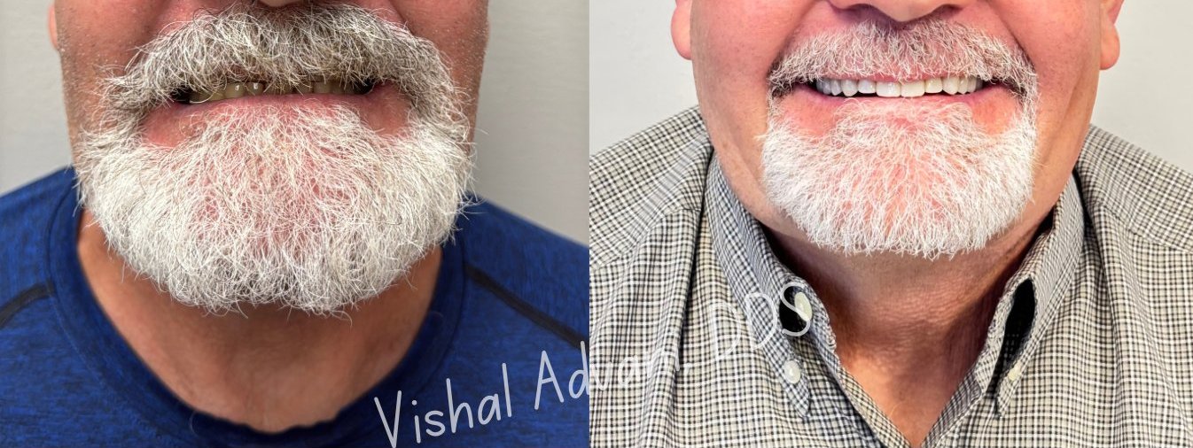
Walk in with a failing front tooth. Walk out with a complete smile — same day. The most aesthetically demanding procedure in implant dentistry, performed with X-Nav dynamic navigation, custom temporary delivery, and obsessive attention to the visible result.
Traditional implant treatment for a failing front tooth involves three separate stages: extraction (with months of healing), implant placement (with months more healing), and finally the crown. The patient lives without a visible front tooth — or with an awkward removable flipper — for most of a year.
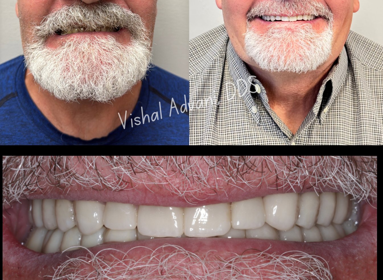
Anterior immediate placement compresses this to a single surgical visit. The failing tooth is extracted, the implant is placed in the same socket, and a custom-shaped temporary crown is delivered the same day. You leave with a complete smile that looks indistinguishable from your natural teeth — even though osseointegration is still occurring beneath the surface.
The technique is demanding. The buccal bone in the anterior maxilla is naturally thin, the gum biotype is often delicate, and the aesthetic margin for error is essentially zero. This is exactly why we use X-Nav dynamic navigation for these cases — sub-millimeter precision is the difference between a result that looks natural and one that looks like an implant.
Beyond the obvious convenience, immediate anterior placement has a real biological advantage: the gum tissue around the extraction site doesn't have time to collapse. The natural papilla (the gum between teeth) and the buccal contour are preserved as they were before extraction.
In a delayed protocol, by contrast, the body begins reabsorbing both bone and soft tissue within weeks of extraction. By the time the implant is placed months later, the gum architecture has changed — and the final crown often requires either grafting to rebuild what was lost, or a compromise on aesthetics.
Time is tissue. Immediate placement preserves what nature already created — and that preserved foundation is what makes the final aesthetic result possible.
A single surgical visit replaces what used to take multiple appointments over many months. Here's exactly what happens.
A 3D CBCT scan and intraoral scan are captured. The implant position is planned digitally, and the temporary crown is designed in advance using your existing tooth as the reference. The temporary is fabricated and ready before you arrive on surgery day.
Under local anesthesia (IV sedation if preferred), the failing tooth is removed using atraumatic technique that preserves the surrounding bone and gum tissue. No flap is raised in most cases — the goal is to disturb as little tissue as possible.
The implant is placed using X-Nav dynamic navigation following the digital plan exactly. Real-time visual confirmation of position, angle, and depth ensures the implant is placed for both biological success and ideal final crown emergence.
The pre-fabricated temporary crown is fitted, adjusted, and delivered. Designed to support the gum tissue and shape it as it heals, the provisional looks and functions like a real tooth from the moment you leave the office.
The implant integrates with the bone while the gum tissue heals around the provisional. The provisional is fully functional during this period — most patients have no functional or aesthetic limitations.
A digital scan captures the matured tissue architecture, and a custom zirconia crown is fabricated and delivered. This is your long-term restoration — designed to look indistinguishable from your natural teeth and last decades.
Most patients with a failing or recently lost anterior tooth are candidates for immediate placement. The decision comes down to your specific anatomy, the condition of the bone surrounding the tooth, and the overall health of the surrounding tissue.
The most common scenario. A tooth needs to come out — typically due to fracture, deep decay, or root resorption — and you need a replacement plan.
The bone on the cheek-side of the tooth must be intact (this is verified with the CBCT scan). For cases with damaged buccal bone, FP1 surgery with socket shield techniques may be a better option.
The teeth on either side should be healthy or stable. If they have their own issues, those are addressed first or as part of a comprehensive plan.
Active infection or significant inflammation at the extraction site may require staged treatment. Most cases that have been managed appropriately are candidates.
Anterior immediate placement is most beneficial for patients for whom the visible appearance matters — anyone with a public-facing job, professional speakers, or anyone who simply doesn't want to wear a gap or flipper.
A complimentary consultation includes a CBCT scan and a digital preview of your predicted aesthetic outcome. For a failing front tooth, the conversation is the most important first step — the rest of the timeline can move quickly.