The most refined approach to single-tooth implant replacement: socket shield, root membrane, partial extraction therapy, pontic shield, and root banking — surgical techniques that preserve your natural tissue architecture so the final result looks indistinguishable from a real tooth.
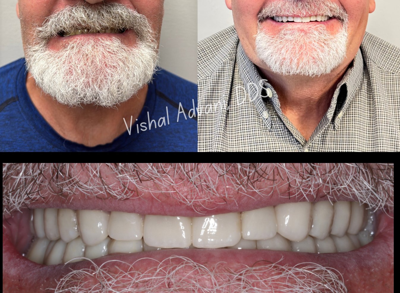
The "FP1" classification — short for Fixed Prosthesis Type 1 — describes a dental implant restoration that replaces only the crown of the missing tooth, with the gum line and surrounding tissue preserved exactly as nature created them. The result is an implant that looks indistinguishable from a real tooth, even when smiling fully.
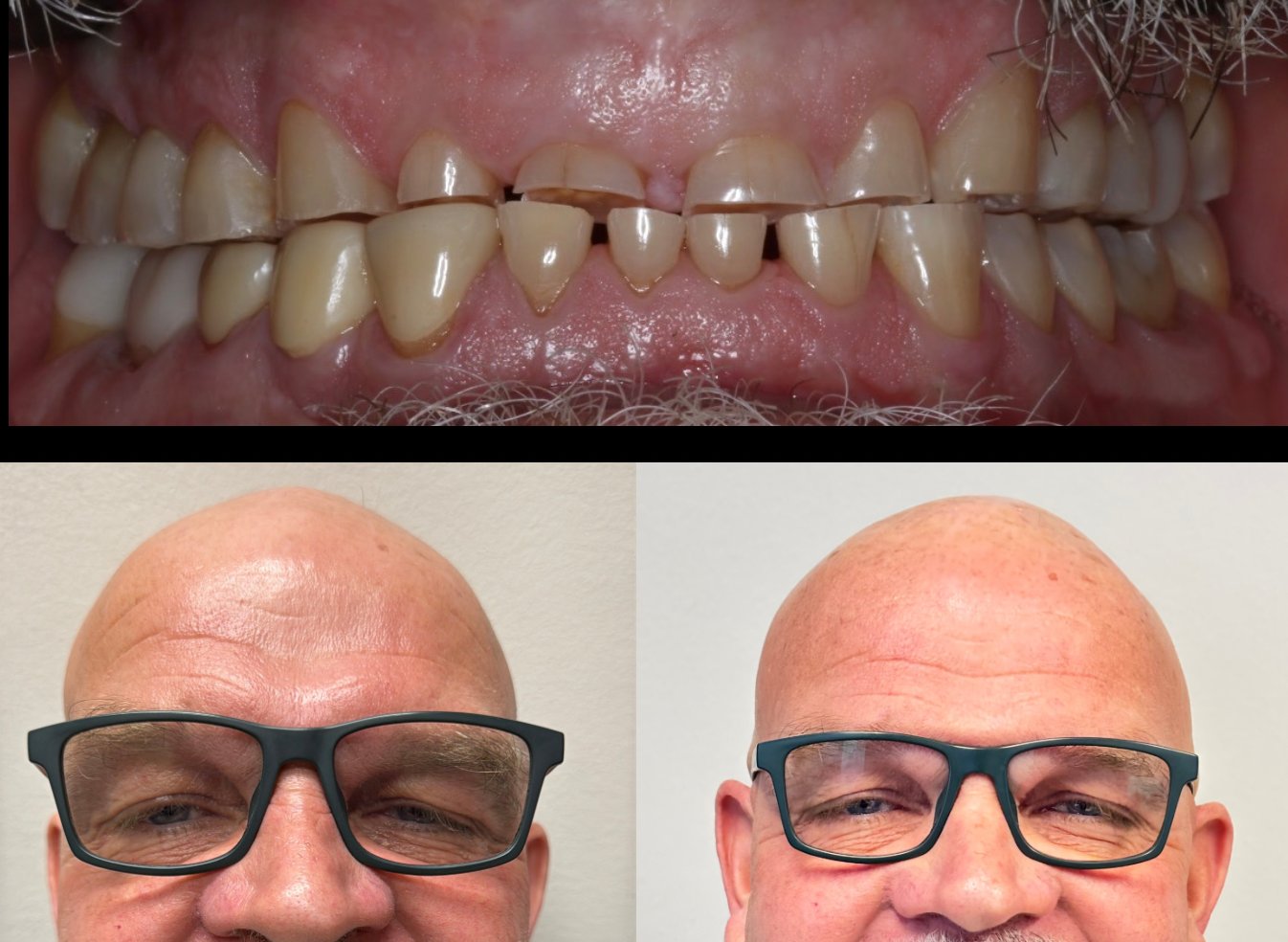
The challenge is that FP1 only works when the bone and soft tissue around the extraction site are preserved. The moment a tooth is removed, the body begins reabsorbing the bone and gum that supported it — and within months, what remains is often inadequate for a true FP1 result. This is why most implant cases default to FP3, where pink prosthetic gum material is added to compensate for lost tissue.
FP1 surgery uses biological preservation techniques at the time of extraction to keep the tissue architecture intact — so when the final crown is placed, the result is a natural smile, not a prosthetic one.
Each of these techniques preserves a different aspect of the natural tissue architecture. The right choice depends on your specific anatomy, the condition of the tooth being replaced, and the aesthetic demands of the case.
A thin slice of the natural tooth root is intentionally retained on the buccal (cheek-side) of the socket during extraction. This shield maintains the periodontal ligament's blood supply and prevents the buccal bone from collapsing — preserving the gum line precisely.
A modification of socket shield that retains a thinner, shaped root remnant — providing the same biological benefit while reducing the technical complexity. Particularly effective in narrow-ridge anterior cases.
An umbrella protocol that combines selective root retention with immediate implant placement. Indicated when the tooth's root structure can serve as a natural scaffold for both the implant and the surrounding tissue.
For cases where a bridge spans an extraction site, a portion of the natural root is preserved beneath the pontic (replacement tooth). This maintains the ridge contour and prevents the typical sunken appearance under bridge pontics.
Strategic preservation of root structure during extraction for patients who may need an implant later. Allows future implant placement without the bone loss that typically follows months or years of an empty socket.
Most real cases use these techniques in combination, tailored to your anatomy. Pre-surgical planning with Blue Sky Plan determines which approach fits your specific situation — and what the predicted aesthetic outcome will be.
FP1 surgery cannot be planned freehand. The margin for error in tissue preservation is too small. Every FP1 case at the Institute begins with Blue Sky Plan — implant planning software that uses your CBCT scan to map the exact position, depth, and angulation of the implant in three dimensions, calibrated against the planned final crown.
The Blue Sky Plan output drives the entire surgical workflow: from where the socket shield will be cut, to where the implant will be placed, to how the same-day provisional will fit. The plan is then executed using X-Nav dynamic navigation — meaning what was planned digitally is what happens surgically, accurate to within fractions of a millimeter.
FP1 is a single-visit surgical procedure with a 3-4 month healing window before the final crown. Here's exactly what to expect at each stage.
3D imaging captures your bone, tooth root, and surrounding anatomy. Blue Sky Plan software is used to design the implant position, the socket shield geometry, and the planned final restoration. You preview the predicted aesthetic result before any surgery happens.
Under local anesthesia (or IV sedation if preferred), the buccal portion of the natural root is retained as a shield while the rest of the tooth is removed. The implant is placed using X-Nav dynamic navigation following the Blue Sky Plan exactly. A custom abutment is connected.
A provisional crown is delivered the same visit so you leave with a complete smile. The provisional is shaped to support the gum tissue and guide its healing into the precise contour planned digitally.
During this period, the implant osseointegrates with the bone while the soft tissue heals around the provisional crown. Most patients have no functional limitations during this phase. Routine check-ins ensure tissue health.
A digital scan captures the exact tissue architecture, and a custom zirconia or e.max crown is fabricated and placed. The result: a tooth that looks indistinguishable from your natural dentition, with no visible metal, no pink acrylic, and a gum line that matches the surrounding teeth precisely.
FP1 surgery is most beneficial when aesthetics matter — typically anterior teeth, or any visible-zone tooth where the result must look indistinguishable from natural dentition. It's also beneficial whenever maximum tissue preservation is desired, regardless of position.
FP1 is the ideal solution when a front tooth needs to be replaced. The visible gum line is preserved, eliminating the risk of an aesthetic compromise.
Patients whose smiles show significant gum tissue benefit most from FP1. With FP3 restorations, the transition between natural and prosthetic gum often becomes visible.
Patients with naturally thin gum tissue are most vulnerable to bone collapse after extraction. Biological preservation prevents this from the start.
For patients who want the best possible aesthetic outcome over the next 20+ years — not just the year after surgery — FP1 preserves the foundation that makes long-term aesthetic stability possible.
A complimentary consultation includes CBCT imaging, a Blue Sky Plan preview of your predicted outcome, and a transparent treatment plan. For patients prioritizing aesthetic outcomes, the conversation alone is worth the visit.